 +1-2404726069 (U.S.)
 0
0Want to control tumor growth in nude mice in real time? Do you want to know the location of cell colonization in mice? Want to know the effect of drug treatment on tumors in vivo? These can be achieved by installing a tracker on the cell, allowing you to control the location and number of cells at any time. This technology is in vivo imaging "detection" technology. So what is in vivo imaging technology?
1. What is in vivo imaging technology?
2. Characteristics of luciferase imaging
3. Application direction of luciferase imaging
4. Experimental example sharing
5. FAQ
6. Product information
7. Regarding reading
1. What is in vivo imaging technology?
As early as 1999, Dr. Weissleder of Harvard University in the United States proposed the concept of molecular imaging, that is, using imaging methods to conduct qualitative and quantitative research on biological processes in vivo at the cellular and molecular levels. In vivo imaging is based on molecular imaging. Through this imaging system, biological processes such as tumor growth and metastasis, the development of infectious diseases, and the expression of specific genes can be observed in living animals.
In vivo, optical imaging of living animals mainly adopts two technologies: bioluminescence and fluorescence. Bioluminescence is the luciferase gene to mark cells or DNA, while fluorescence technology uses fluorescent reporter genes such as green fluorescent protein and red fluorescent protein and fluorescence such as FITC, Cy5, and Cy7. Elements and quantum dots (QD) for labeling. Mammalian bioluminescence generally integrates the firefly luciferase gene (composed of 554 nucleotides, about 50KD), that is, the luciferase gene, into the chromosomal DNA of the expected observation cell to express luciferase. Then cultivate a cell line that can stably express luciferase, and when the cells divide, differentiate, and transfer, the luciferase will also continue to express stably. Genes, cells, and live animals can all be tagged with the luciferase gene. Luciferase is a kind of enzyme that can catalyze substrates to produce bioluminescence. Luciferases from different sources have their characteristics and can catalyze substrates to emit different colors of light. Among them, firefly luciferase has high sensitivity and a wide linear range of 7~8 orders of magnitude. It has become the most commonly used mammalian cell reporter gene. The luciferase reporter plasmid was transferred into the cells, and its substrate luciferin was added to incubate the cells. In the presence of ATP, O2, and magnesium ions, luciferase could oxidize the luciferin substrate to produce a visible light reaction. Realize "one-time 'tracker' installation, and track and detect at any time". In addition to firefly luciferase, renilla luciferase is sometimes used. The substrates of the two are different, the substrate of the former is D-luciferin, and the substrate of the latter is coelenterazine. The light emitting wavelengths of the two are different, the range of the light wavelength emitted by the former is 540-600nm, and the range of the light wavelength emitted by the latter is 460-540nm. The light emitted by the former is easier to pass through tissues, while the latter is metabolized faster in the body, and its specificity is not as good as the former. Therefore, not most in vivo experiments using firefly luciferase as a reporter gene.
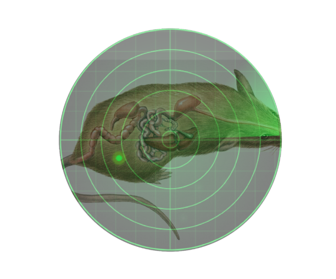
Figure 1. Localization of luciferase labeled cells
The optical principle of bioluminescence: light will be scattered and absorbed when propagating in mammalian tissues, and photons will be refracted when encountering cell membrane and cytoplasm, and different types of cells and tissues have different characteristics of absorbing photons. Hemoglobin is the main cause of the absorption of visible light in the body and can absorb most of the blue-green band of visible light. But in the red light band of visible light greater than 600nm, the absorption of hemoglobin is very small. Therefore, a large amount of light can pass through tissue and skin to be detected in the reddish region. At least a few hundred subcutaneous cells can be detected using live animal bioluminescent imaging technology. However, depending on the depth of the light source in the mouse, the minimum number of cells that can be seen varies. Generally speaking, for every increase of 1cm, the luminous intensity attenuates by 10 times, and the attenuation is more for tissues and organs rich in blood, and less attenuation for tissues and organs adjacent to bones. In the case of the same depth, the detected luminous intensity has a significant linear relationship with the number of cells, and the detected light intensity can be quantified by the instrument to reflect the number of cells.
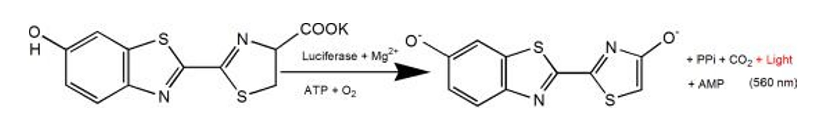
Figure 2. The luminescent principle of luciferase and luciferin potassium salt reaction
Different from bioluminescence, fluorescence technology uses fluorescent reporter genes or fluorescent dyes (including new nano-labeling materials such as fluorescent quantum dots) for labeling. Using fluorescence from reporter genes, fluorescent proteins, or dyes, a biological light source in vivo can be created. Bioluminescence is autofluorescence in animals without an excitation light source, while fluorescence requires excitation by an external excitation light source before it can be detected by the imaging system. Fluorescent labels are widely used, including animals, cells, microorganisms, antibodies, drugs, nanomaterials, etc.
2. Characteristics of luciferase imaging
◎ no radiation, almost harmless to organisms.
◎ bioluminescence without excitation light source.
◎ high sensitivity, hundreds of cells can be detected.
◎ good penetrability, 3-4cm tissue depth can still be detected.
◎ high signal-to-noise ratio, strong fluorescence signal, and good anti-interference.
3. Application direction of luciferase imaging
3.1 Tumor growth
In the tumorigenesis experiment in nude mice, the tumor growth was observed in real time without invasion, and there was no need to strip the tumor for measurement.
3.2 Oncology drugs
The influence of administration on tumor growth or metastasis was detected, and the fluorescein substrate could be eliminated within 3 hours, without interference with the drug experiment.
3.3 Cell localization
The localization and distribution of foreign cells in animals were detected.
3.4 Gene expression regulation
The target gene or the promoter of the target gene was fused with the luciferase gene to detect gene expression changes during drug treatment or the course of the disease.
3.5 Stem cell research
Monitoring the transplantation, survival, and proliferation of stem cells; Tracing the distribution and migration of stem cells in vivo.
4. Experiment example sharing

Figure 3. in vivo imaging detection of the therapeutic effect of CAR-MUC1 T/CAR-MUC1-IL22 T cells on tumor formation by subcutaneous injection of HN4 cells in mice[1].
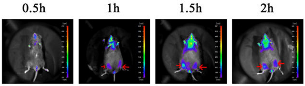
Figure 4. After HUC-MSCs cells were injected into mouse skeletal muscle, the localization of cells was detected by in vivo imaging (marked by red arrow)[2].
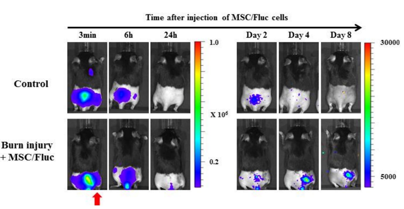
Figure 5. The ability of in vivo imaging to detect the migration of mesenchymal stem cells (MSC) to burn sites. Mesenchymal stem cells (MSC/FLuc) were injected intravenously into the mouse back burn model. Four days after injection, bioluminescence signals appeared at the injured site of the burn wound, and then gradually decreased (the red arrow indicates the burn site)[3].
5. FAQ
Q1: Compared with traditional technology, what are the advantages of bioluminescence imaging technology?
Compared with traditional technology, this technology is more sensitive than the traditional methods in the research of tumor metastasis, gene therapy, epidemiology, stem cell tracing, leukemia, and other related research. It can also quickly and intuitively study the pathogenesis and drug screening of related diseases through a series of transgenic animal disease models.
Q2: How to label stem cells with the luciferase gene?
The constitutively expressed genes can be labeled to make transgenic mice, and the stem cells are labeled. The hematopoietic stem cells are taken from the mouse's bone marrow and transplanted into another mouse's bone marrow. This technology can be used to trace the proliferation, differentiation, and migration of hematopoietic stem cells in the body. Another method is to label stem cells with lentivirus.
Q3: How long is it appropriate to test after fluorescein injection, and how long can the luminescence last?
Generally, the fluorescence signal reaches the strongest stable period after intraperitoneal injection for 10-15min and begins to decay after 20-30min. After 3h, the fluorescein is eliminated and the luminescence disappears completely.
Q4: How to inject fluorescein into mice? What is the difference between injection methods?
Fluorescein can be injected into mice by intraperitoneal injection or tail vein injection. It can spread to the whole body of mice in about 1 min. In most cases, the concentration of fluorescein is 150 mg/kg. For 20 g mice, about 3 mg of fluorescein can be used. For intraperitoneal injection, the diffusion is slow, the initial luminescence is slow, and the continuous luminescence time is long. For the tail vein injection of fluorescein, it diffuses quickly and starts to emit light quickly, but the duration of luminescence is short.
6. Product information
Yeasen is a biotechnology company engaged in the research, development, production, and sales of three major biological reagents: molecules, proteins, and cells. The products provided by Yeasen are as follows.
Table 1. Product information
| Product information | Product code | Specifications |
| D-Luciferin, Sodium Salt | 40901ES01/02/03/08/10 | 0.1/0.5/1/5/10 g |
| D-Luciferin, Potassium Salt | 40902ES01/02/03/08 | 0.1/0.5/1/5 g |
| D-Luciferin Firefly, Free Acid (Inquire) | 40903ES01/02/03 | 0.1/0.5/1 |
| Coelenterazine h (Inquire) | 40906ES02/03/08 | 0.5/1/5 mg |
| Ready To Use Coelenterazine h (Inquire) | 40907ES10 | 10 vials |
| Dual Luciferase Reporter Gene Assay Kit (Inquire) | 11402ES60/80 | 100/1000T |
| Luciferase Reporter Gene Assay Kit (Inquire) | 11401ES60/76/80 | 100/500/1000T |
| VDR (Vitamin D Receptor) Luciferase Reporter Plasmid (Inquire) | 11502ES03 | 1μg |
| STAT1 Luciferase Reporter Plasmid (Inquire) | 11504ES03 | 1μg |
7. Regarding reading
A new generation of luciferase reporter gene detection system——Easier, more sensitive, more precise
