Products in stock in the United States can be delivered within 2 days.
 +1-2404726069 (U.S.) +86-027-65528241 (Outside U.S.)
  overseas@yeasen.com shop website:https://yeabio.com
 0
0Blasticidin S (10mg/ml in Solution)

Product Description
Blasticidin S is a nucleoside antibiotic from Streptomyces griseochromogenes that specifically inhibits protein synthesis in prokaryotes and eukaryotes by interfering with the formation of peptide bonds in the ribosome. Blasticidin S is used to screen transfected cells carrying BSR or BSD genes. Blasticidin has a rapid and potent mode of action, causing rapid cell death at very low antibiotic concentrations.
Cat#60218ES10, Cat#60218ES50, Cat#60218ES60 are provided in liquid form at a concentration of 10 mg/mL (HEPES buffer, pH 7.5).
Cat#60218ES80 is provided in powder form and can be prepared as a 5-10 mg/mL stock solution with sterile water or buffer.
Resistance gene: There are three kinds of pyrintin tolerance genes that have been cloned and sequenced, one of which is isolated from Streptoverticillum sp. The other two are deaminase genes, including Bsr isolated from Bacillus cereus and Bsd isolated from Aspergillus terreus. Bsr and Bsd genes are the most commonly used screening markers for gene transfer experiments in mammalian and plant cells, and can also be used in E. coli.
Product Properties
|
CAS No. |
3513-03-9 |
|
Molecular formula |
C17H26N8O5·HCl |
|
Molecular weight |
458.9 g/mol |
|
Purity |
≥95% |
|
Structure |
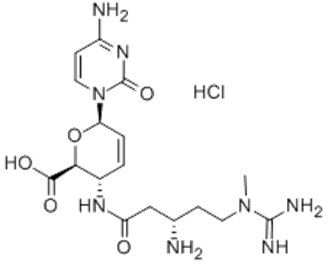 |
Shipping and Storage
The product is shipped with an ice pack and can be stored at -20℃. Powder lasts for 3 years and liquid lasts for 1 year. Avoid repeated freezing and thawing.
Application of concentration
1) Escherichia coli
E. coli is slightly less sensitive to Blasticidin S, but transformants are tolerant to Blasticidin S and can be screened in low-salt LB medium (pH 8) at concentrations ranging from 50-100 μg/mL Blasticidin S. High pH can increase the activity of Blasticidin S.
2) Mammalian cell
The working concentration of Blasticidin S in mammalian cells ranges from 1 to 50 μg/mL. Initial experiments suggest using kill curves to determine the optimal concentration for use.
Table 1. Recommended working concentrations for mammalian cells
|
Cell |
species |
Tissues |
Medium |
Concentration of Blasticidin S (μg/mL) |
|
HeLa |
Human |
Uterus |
DMEM |
3-10 |
|
293 |
Human |
Kidney |
DMEM |
3-10 |
|
B16 |
Mouse |
Melanoma |
RPMI |
3-10 |
|
PC1.0 |
Hamster |
Adenocarcinoma |
RPMI |
10-30 |
Instructions (Mammalian stable strain screening)
Blasticidin S is usually used at a concentration of 10 μg/mL. Plasmids carrying BSR or BSD genes were transfected into cells and incubated in a normal growth medium containing Blasticidin S for the selection of stably transfected cell lines.
1) 48 h after transfection, cells were subcultured with a fresh medium containing a suitable concentration of Blasticidin S (Note: Antibiotics work best when the cells are actively dividing. If the cell density is too high, the antibiotic efficiency will be reduced. It is best to have no more than 25% coverage when cells are divided.)
2) Remove the culture medium every 3-4 days and add fresh culture medium containing antibiotics.
3) Colony formation was detected after 7 days. Colony formation may take an additional week or more depending on host cell type and transfection/screening efficiency.
4) Transfer 5-10 resistant clones to a 35 mm cell dish and add a selective medium to maintain the culture for 7 days. It was subsequently tested with a cytotoxicity assay.
Cautions
1. For your safety and health, please wear lab coats and disposable gloves for operation.
2. For research use only!
[1] Xiao J, Xiong Y, Yang LT, et al. POST1/C12ORF49 regulates the SREBP pathway by promoting site-1 protease maturation. Protein Cell. 2021;12(4):279-296. doi:10.1007/s13238-020-00753-3(IF:10.164)
[2] Wei W, Li Y, Wang C, et al. Diterpenoid Vinigrol specifically activates ATF4/DDIT3-mediated PERK arm of unfolded protein response to drive non-apoptotic death of breast cancer cells [published online ahead of print, 2022 Jun 1]. Pharmacol Res. 2022;182:106285. doi:10.1016/j.phrs.2022.106285(IF:7.658)
[3] Qian F, Hu Q, Tian Y, et al. ING4 suppresses hepatocellular carcinoma via a NF-κB/miR-155/FOXO3a signaling axis. Int J Biol Sci. 2019;15(2):369-385. Published 2019 Jan 1. doi:10.7150/ijbs.28422(IF:6.582)
[4] Li Q, Yu D, Yu Z, et al. TIPE3 promotes non-small cell lung cancer progression via the protein kinase B/extracellular signal-regulated kinase 1/2-glycogen synthase kinase 3β-β-catenin/Snail axis. Transl Lung Cancer Res. 2021;10(2):936-954. doi:10.21037/tlcr-21-147(IF:6.498)
[5] Li Y, Fu Y, Hu X, et al. The HBx-CTTN interaction promotes cell proliferation and migration of hepatocellular carcinoma via CREB1. Cell Death Dis. 2019;10(6):405. Published 2019 May 28. doi:10.1038/s41419-019-1650-x(IF:5.959)
[6] Li X, Wu A, Han C, et al. Bone marrow-derived mesenchymal stem cells in three-dimensional co-culture attenuate degeneration of nucleus pulposus cells. Aging (Albany NY). 2019;11(20):9167-9187. doi:10.18632/aging.102390(IF:5.515)
[7] Yue C, Jin H, Zhang X, et al. Aucubin prevents steroid-induced osteoblast apoptosis by enhancing autophagy via AMPK activation. J Cell Mol Med. 2021;25(21):10175-10184. doi:10.1111/jcmm.16954(IF:5.310)
[8] Wu J, Lv Q, He J, et al. MicroRNA-188 suppresses G1/S transition by targeting multiple cyclin/CDK complexes. Cell Commun Signal. 2014;12:66. Published 2014 Oct 11. doi:10.1186/s12964-014-0066-6(IF:4.672)
[9] Huang H, Zou X, Zhong L, et al. CRISPR/dCas9-mediated activation of multiple endogenous target genes directly converts human foreskin fibroblasts into Leydig-like cells. J Cell Mol Med. 2019;23(9):6072-6084. doi:10.1111/jcmm.14470(IF:4.658)
[10] Huang H, Zhong L, Zhou J, et al. Leydig-like cells derived from reprogrammed human foreskin fibroblasts by CRISPR/dCas9 increase the level of serum testosterone in castrated male rats. J Cell Mol Med. 2020;24(7):3971-3981. doi:10.1111/jcmm.15018(IF:4.486)
[11] Wang X, Du A, Yu G, Deng Z, He X. Guanidine N-methylation by BlsL Is Dependent on Acylation of Beta-amine Arginine in the Biosynthesis of Blasticidin S. Front Microbiol. 2017;8:1565. Published 2017 Aug 22. doi:10.3389/fmicb.2017.01565(IF:4.076)
[12] Zhao Y, Ye X, Chen R, et al. Sirtuin 7 promotes non‑small cell lung cancer progression by facilitating G1/S phase and epithelial‑mesenchymal transition and activating AKT and ERK1/2 signaling. Oncol Rep. 2020;44(3):959-972. doi:10.3892/or.2020.7672(IF:3.906)
[13] Zhang M, Liu X, Wang Q, et al. NDRG2 acts as a PERK co-factor to facilitate PERK branch and ERS-induced cell death. FEBS Lett. 2017;591(21):3670-3681. doi:10.1002/1873-3468.12861(IF:3.623)
[14] Wang XF, Tian Q, Qin WB, et al. Histone H3 methylation orchestrates transcriptional program in mouse spermatogenic cell line. J Reprod Dev. 2020;66(3):223-230. doi:10.1262/jrd.2019-139(IF:1.735)
Catalog No.:*
Name*
phone Number:*
Lot:*
Email*
Country:*
Company/Institute:*

